Wednesday, June 3, 2009

Mutation in filaggrin gene trigger allergies


Eczema can cause the skin to become very red and itchy


Breakthrough of genetic defect in mice enable scientist to identify what factor is responsible for the rapid increase in allergic today.

Scientists in Dundee, Ireland and Japan discover the filaggrin defect in mice which causes allergic inflammation , similarly like what happen in human eczema.They found out that the filaggrin gene is very important since this gene enable skin to produce protective barrier.So, this will block the allergens from entering the body,thus preventing allergic formation.

Scientist pointed out that about one in five children in Britain and other westernised nations suffer from eczema.

Eczema cause irritating patch of sore skin,while in extreme cases ,extensive areas may become inflamed and unbearably itchy. So, the children with this condition are very susceptible to get allergic conditions, like asthma , fever and many more.

The finding indicates that many cases of eczema are induced by genetic mutation in filaggrin gene.

The breakthrough of genetic defect of filaggrin gene enable enable scientist to develop therapies for eczema and other allergies by suppressing the defective filaggrin gene.Now, scientists are still looking for the drugs or treatments that aim at the filaggrin gene and hopefully they will find the cure for these disease in recent time.

prepared by

42101765...tuc

REFERENCE…

Mouse Gene Aids Allergy Research 2009, BBC News, 6 April, viewed 28 May 2009 at

http://news.bbc.co.uk/2/hi/uk_news/scotland/tayside_and_central/7985307.stm%3e

Link to original source of article…

http://news.bbc.co.uk/2/hi/uk_news/scotland/tayside_and_central/7985307.stm


Multiple Hereditary Exostoses (MHE)


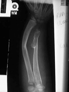
Multiple Hereditary Exostoses is an inherited disorder of bone growth. MHE can be referred to by various names such as Heredity Multiple Exostoses, Hereditary Multiple Osteochondromata, Multiple Carthaginous Exostoses, etc. People who have MHE grow exostoses, or bony bumps, on their bones which can vary in size, location and number depending on the individual. Although any bone can be affected, the long bones (legs, arms, fingers, toes), pelvis and shoulder blades are the most common, while the face and skull are generally unaffected. Boys and girls can both be affected. Older literature claimed that boys were more severely troubled by MHE, but bigger series of patients studied recently do not support this theory.

MHE is a condition that is passed by the genes of the affected parent to their children. It is called an ~autosomal dominant~ disorder which means that if one parent has the condition, chances are fifty percent that any child could also develop MHE. Occasionally, a patient will develop multiple exostoses with no previous family history of MHE. This situation is described as a spontaneous mutation meaning a genetic problem arose in that person without being inherited from a parent. Recently chromosomes (the packages that carry genes) 8, 11 and 19 have all been shown to be locations where the genetic information for MHE comes from. Some researchers feel there actually may be different types of MHE each caused by different genes at these locations.

An exostosis is a bone growth that is abnormal or different from the underlying architecture of the bone. These "abnormal growths" are not cancer, They are benign. Sometimes doctors refer to exostoses as "tumors" which like exostose is a general term meaning abnormal growth. It is important to remember that not all "tumors" are cancer. Most tumors, like the exotoses of MHE, are benign. Exostoses start near the growth centers of bones which are near the ends of the bones, which is why bumps grow near the joints. They can be rounded or sharp and continue to grow while a child is growing. When a person is full grown, exostoses also stop growing.

MHE can be troublesome because the exostoses grow near the growth centers of the bone, they can make the growth center grow poorly, or only part of it grow poorly. This makes a lot of people with MHE somewhat shorter than average or have bowed arms or legs. Often, the forearm will bow out toward the lime finger, or the legs can become knock kneed. This is frequently concerning, but function is often normal though cosmetically, the bowing can be very troubling. Sometimes folks with MHE get stiff, especially in the elbows and hips, usually because their exostoses block some of their motion. While children are growing, exostoses can be painful. They seem to be very sensitive to getting bumped. Kids often develop exostoses on the inside of their knees and these can hit together when they run, which hurts!

Sometimes exostoses grow near nerves or tendons and press on them. In these cases, they often need to be removed so they won't damage the structure laying over them.

The most frightening complication of MHE is also one of the most uncommon. Rarely (less than 1% of the time), the benign exostoses of MHE can become a malignant tumor called chondrosarcoma. This happens almost always after adulthood when skeletal growth has ceased. Usually, patients who develop chondrosarcoma are in their 20's to 50's. If a person with MHE notices that an exostoses is getting bigger or painful after they have stopped growing they should get to their doctor! Growth and pain are two important warning signs that a benign tumor has become malignant. Chondrosarcoma is very rare, but it is something MHE families must know about.

According to this article, some people with MHE never require any treatment. They learn to compensate for deformity or decreased range of motion so they function normally. When deformity does occur, it often happens so slowly that the patient can compensate for it well, while others may require surgery to help them.

If an exostoses is painful, pressing on an important structure, cosmetically unattractive or if easily bumped, it can be surgically removed. Once removed, exostoses can reoccur (about 20 - 50% of the time), but may not regrow to a size large enough to be symptomatic. Removal itself is usually a fairly small procedure; some are removed without ever staying overnight in the hospital.

If an exostoses causes a growth abnormality, like bowing, sometimes just removing the exostoses early enough will allow the bone to straighten itself out and remodel as the child grows. Some bowing is so severe that not only must the exostoses be removed, but also the bone must be straightened. This can be done by either cutting the bone, straightening it and then holding it in place while it heals or if the child is still growing by changing the rate of growth on one side of the growth plate. Currently there are several options and your doctor should be able to explain them to you.

If an exostoses does become malignant and turn into a chondrosarcoma then it must be removed. A specialist in orthopedics and bone tumors would be required to help with this.

Scientists throughout tho world have demonstrated the genes for MHE are found on three different chromosomes. This leads to the belief that MHE is caused by at least three different genes with one or more on each chromosomes. It is known that the genes are located on chromosome numbers 8, 11 and 19 with number 8 being the most common location found.

Continuing research of the genes and how the proteins encode for them will give tremendous insight into the growth of cells. This information is important since MHE is a problem with the growth of cells.

Understanding the gene and the function of its protein might eventually provide the knowledge leading to actual treatment. The gene mapping studies will serve as the basis for the testing of children at risk for MHE. At the time of this publication, the availability of such testing is limited to a research setting. However, your physician could be equipped to perform this test in the near future. Information from this test could lead to the prevention of the development of exostoses and their complications.

Posted by:

Aini Syahida binti Mat Yassim

42101167

Reference

http://www.wheelessonline.com/images/med2.jpg

http://www.radix.net/~hogue/mhe.htm

Mice Carrying a 'Humanised' Gene Teach us About Speech and Language

Jonathan Whitburn
42052531

Wolfgang Enard of the Max-Planck Institute for Evolutionary Anthropology and his team have been studying mice carrying a "humanised gene"; in humans, this gene is believed to be responsible for speech and language. Enard and his team are interested in this gene in particular because it is one of the important genomic differences between humans and primates; what make the gene different are two amino acid substitutions in the gene FOXP2. Previous studies have shown that this gene has undergone positive selection, but only in humans because the gene wasn’t fixed until after the lineage broke between humans and chimpanzees. Important aspects of speech and language are thought to be the result of the evolutionary change. Mice, which have been used for mainly studies of disease, are now becoming tools for studying our history; that is, Enard and his team have been studying the effects of the humanised FOXP2 gene in mice.

The study showed that brain circuits, linked to human speech in previous studies, are present in mice containing the human FOXP2 gene. Enard and co. found that there are also qualitative differences in ultrasonic vocalisations between baby mice with the human gene and without; the vocalisations were brought about by placing the pups outside the mother's nest. There is not enough information, however, regarding mouse communication to look further into the qualitative changes. It is known that FOXP2 is active in other cells in the body, but in the mice, no other physical changes were recorded; the mice appeared to be healthy. Enard's team speculated that this gene could cause fine-tuning of motor control in the lungs, larynx, tongue and lips that cause articulation in speech.

Source:
Cathleen Genova
Cell Press
Original Article Title: ‘Humanized’ Mice Speak Volumes, retrieved from:
http://www.medicalnewstoday.com/articles/151866.php

Genetically Modified Cells Migrate To Brain And Treat Neurodegeneration In St. Jude Model

Name: Joanne Tham (s4209695)


This article is about the effect of a genetically modified gene that has the ability to cure patients from a fatal disease known as Lysosomal Storage disease (LSDs), by being able to transport the desired drug to dying neural cells in the brain.

This cure was discovered by St.Jude researchers who were trying to treat a type of LSDs known as GM1--Gangliosidosis by using Bone Marrow Cells (BMC) and inserting an enzyme that breaks down fat molecules GM1. GM1 is an essential component in the brain, however patients who are suffering from GM1--Gangliosidosis lack the enzyme beta-glycosidase. This results in an increased concentration of GM1 which damages the cell and causes it to die. The function of BMC is that it produces a wide variety of cells with different functions, one example being the immune cells known as monocytes. This gene was then genetically modified and inserted into the a laboratory model. The results showed that the monocytes migrated toward the site lacking in beta-glycosidase, the enzymes released were then used by the cells to break down excess GM1 which help to prevent the buildup of more GM1.

The migration of the monocytes were because they followed the gradient-increasing concentration signals being produced by the cells adjacent to the dying neurons known as chemokines until they reached the dying neurons. In normal circumstances, the immune cells would cause greater problems to the neurons as they made them became inflamed. However, because these cells were genetically modified monocytes, it helped to restore the beta-glycosidase activity which reduced the levels of dying neurons as well as chemokines.

In conclusion, if the BMCs were able to be successfully modified, this would be an effective form of therapy. The genetic modification of the BMCs would be able to produce unlimited amounts of the modified monocytes, hence ensuring that there would always be sufficient enzymes to reduce the activity of beta-glycosidase. In addition, if the cells were able to be replicated by the same patient after they were modified, this would also help to get rid of complications in finding a suitable donor.


Reference List:

St. Jude Children's Research Hospital (2005) “Genetically Modified Cells Migrate To Brain And Treat Neurodegeneration In St. Jude Model” Available at: http://www.sciencedaily.com/releases/2005/07/050718005942.htm Viewed on 12/5/09

How The swine flu virus will be created


The H1N1 is spreading fear across the Planet: The following is a short summary of how scientists plan to make a vaccine for this virus

There are 2 methods: The first involves drilling a hole in a hens egg and injecting a small amount of the virus into it. The egg is then left to incubate and after a few days there will be enough of the virus to begin phase 2. Here scientists use reverse genetics and take the surface protein of H and N from the H1N1 virus and mix them with a common lab virus known as PR8. This creates a harmless hybrid which can be used as a vaccine.

The second method involves the same first step with the hens egg. Except this time both the virus and the PR8 virus are injected into the egg allowing a new recombinant strain to be created due to the natural re-assortment of their genes.

The vaccine will trick the immune system of the patient into thinking it has the virus, thereby forcing it to create antibodies for it, so that when the actual virus strikes the antibodies are ready and able to protect the person.

it should take a few months for this vaccine to be commercially available

Reference: http://news.bbc.co.uk/1/hi/health/8029917.stm - accessed 3/6/09

New Hopes for Early and Faster Detection of HCM Cardiac Disease
Hypertrophic cardiomyopathy (HCM) is a familial cardiac disease caused by a variety of mutant genes encoding protein components of the cardiac sarcomere, transmitted to each consecutive generation as an autosomal dominant trait with variable penetrance and heterogeneous clinical expression [1]. The traditional approach to screening relatives in hypertrophic cardiomyopathy (HCM) families has been a long arduous process of echocardiography and electrocardiogram ECG on a 12- to 18-month basis, usually beginning at about age 12 years [1]. Until recent discoveries highlighted that the onset of left ventricle hypertrophy can occur well into adulthood; practitioners have assumed that if at the age of twelve that there were no results from the scans that HCM was present then is was most probably absent. Recent advancements in genetics have utilised genetic markers for stratification of sudden death risk and other adverse consequences of HCM. The developments have also highlighted the capability of achieving a diagnosis of HCM with deoxyribonucleic acid (DNA)-based laboratory methods; which is irrefutable and has led to enhanced recognition of the HCM disease state, and consequently to more complete definition of its broad clinical spectrum, as well as providing practical insights into appropriate genetic counselling [1].
Intense studies conducted on the HCM genotype have identified more than 200 mutants responsible for the disease. Two of the HCM-causing mutant genes, beta-myosin heavy chain and myosin-binding protein C, appear to predominate in frequency. The other eight genes appear to account for far fewer cases of HCM and include troponin T and I, regulatory and essential myosin light chains, titin, alpha-tropomyosin, alpha-actin, and alpha-myosin heavy chain. This discovery of the causes of HCM has helped to quicken the screening process for the diseases, leading the way for future studies in finding a cure.

Removal of One Protein Can Slow Atherosclerosis Progression

May 11 2009, 11:24 AM EST

Removal of One Protein Can Slow Atherosclerosis Progression
GEN News Highlights

Removing a single protein prevents early damage in blood vessels from triggering a later-stage, frequently lethal complication of atherosclerosis, according to researchers at the University of Rochester School of Medicine and Dentistry. By eliminating the gene for cyclophilin A (CypA) from a strain of mice, they were able to provide complete protection against abdominal aortic aneurysm (AAA), according to a study in Nature Medicine.

AAA is a progressive outward dilation of the aorta under the stress of blood pressure due to a breakdown in the vessel's structural integrity. It also shares vital biochemical pathways with atherosclerosis.

“It is extremely unusual for the removal of one protein to provide absolute protection, but it makes perfect sense, because cyclophilin A promotes three of the most destructive forces in blood vessels: oxidative stress, inflammation, and matrix degradation,” explains Bradford C. Berk, M.D., Ph.D., professor of medicine within the Aab Cardiovascular Research Institute at the university, and senior author of the study.

Among the hormones best known to stimulate oxidative stress in blood vessels is angiotensin II. It has also been seen to have a role in matrix degradation and inflammation. The current study sought to answer whether angiotensin II can achieve these disease-causing effects if CypA is not there to pass on its message.

To clarify the role of CypA, the team engineered mice to no longer produce apolipoprotein E, which increased their cholesterol levels and made them prone to atherosclerosis. From this original line, the team further engineered one group with no CypA, another with extra CypA, and compared both to normal mice as all were treated for a month with angiotensin II.

Mice lacking CypA saw greater than 75% decreases in ROS production, MMP activation, and inflammatory cell influx compared to normal mice. MMPs, or matrix metalloproteinases, degrade the matrix structure of the vessel wall. Additionally, they noticed increases in these elements for mice with extra CypA. Angiotensin II treatment also dramatically increased expression of cytokines, unless CypA was missing.

The team also found high CypA levels in the rupture-prone vessels of humans with AAA, and that major drugs like statins reduce CypA levels, which may partly explain their benefit.

The investigators thus propose that ROS generated via angiotensin II trigger CypA secretion from smooth muscle cells in vessel walls. Once outside the cell, CypA docks into CypA receptor proteins on the same cells to increase ROS production in a vicious cycle.

The scientists are now searching for the specific CypA receptors that if interfered with, would shut down ROS production, CypA secretion, MMP activation, and inflammatory cell recruitment in AAA. They are also working to complete a study that will confirm CypA deficiency significantly slows the progression of atherosclerosis.