Wednesday, June 3, 2009

Mutation in filaggrin gene trigger allergies


Eczema can cause the skin to become very red and itchy


Breakthrough of genetic defect in mice enable scientist to identify what factor is responsible for the rapid increase in allergic today.

Scientists in Dundee, Ireland and Japan discover the filaggrin defect in mice which causes allergic inflammation , similarly like what happen in human eczema.They found out that the filaggrin gene is very important since this gene enable skin to produce protective barrier.So, this will block the allergens from entering the body,thus preventing allergic formation.

Scientist pointed out that about one in five children in Britain and other westernised nations suffer from eczema.

Eczema cause irritating patch of sore skin,while in extreme cases ,extensive areas may become inflamed and unbearably itchy. So, the children with this condition are very susceptible to get allergic conditions, like asthma , fever and many more.

The finding indicates that many cases of eczema are induced by genetic mutation in filaggrin gene.

The breakthrough of genetic defect of filaggrin gene enable enable scientist to develop therapies for eczema and other allergies by suppressing the defective filaggrin gene.Now, scientists are still looking for the drugs or treatments that aim at the filaggrin gene and hopefully they will find the cure for these disease in recent time.

prepared by

42101765...tuc

REFERENCE…

Mouse Gene Aids Allergy Research 2009, BBC News, 6 April, viewed 28 May 2009 at

http://news.bbc.co.uk/2/hi/uk_news/scotland/tayside_and_central/7985307.stm%3e

Link to original source of article…

http://news.bbc.co.uk/2/hi/uk_news/scotland/tayside_and_central/7985307.stm


Multiple Hereditary Exostoses (MHE)


Multiple Hereditary Exostoses is an inherited disorder of bone growth. MHE can be referred to by various names such as Heredity Multiple Exostoses, Hereditary Multiple Osteochondromata, Multiple Carthaginous Exostoses, etc. People who have MHE grow exostoses, or bony bumps, on their bones which can vary in size, location and number depending on the individual. Although any bone can be affected, the long bones (legs, arms, fingers, toes), pelvis and shoulder blades are the most common, while the face and skull are generally unaffected. Boys and girls can both be affected. Older literature claimed that boys were more severely troubled by MHE, but bigger series of patients studied recently do not support this theory.

MHE is a condition that is passed by the genes of the affected parent to their children. It is called an ~autosomal dominant~ disorder which means that if one parent has the condition, chances are fifty percent that any child could also develop MHE. Occasionally, a patient will develop multiple exostoses with no previous family history of MHE. This situation is described as a spontaneous mutation meaning a genetic problem arose in that person without being inherited from a parent. Recently chromosomes (the packages that carry genes) 8, 11 and 19 have all been shown to be locations where the genetic information for MHE comes from. Some researchers feel there actually may be different types of MHE each caused by different genes at these locations.

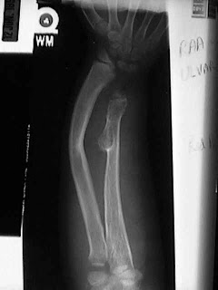
An exostosis is a bone growth that is abnormal or different from the underlying architecture of the bone. These "abnormal growths" are not cancer, They are benign. Sometimes doctors refer to exostoses as "tumors" which like exostose is a general term meaning abnormal growth. It is important to remember that not all "tumors" are cancer. Most tumors, like the exotoses of MHE, are benign. Exostoses start near the growth centers of bones which are near the ends of the bones, which is why bumps grow near the joints. They can be rounded or sharp and continue to grow while a child is growing. When a person is full grown, exostoses also stop growing.

MHE can be troublesome because the exostoses grow near the growth centers of the bone, they can make the growth center grow poorly, or only part of it grow poorly. This makes a lot of people with MHE somewhat shorter than average or have bowed arms or legs. Often, the forearm will bow out toward the lime finger, or the legs can become knock kneed. This is frequently concerning, but function is often normal though cosmetically, the bowing can be very troubling. Sometimes folks with MHE get stiff, especially in the elbows and hips, usually because their exostoses block some of their motion. While children are growing, exostoses can be painful. They seem to be very sensitive to getting bumped. Kids often develop exostoses on the inside of their knees and these can hit together when they run, which hurts!

Sometimes exostoses grow near nerves or tendons and press on them. In these cases, they often need to be removed so they won't damage the structure laying over them.

The most frightening complication of MHE is also one of the most uncommon. Rarely (less than 1% of the time), the benign exostoses of MHE can become a malignant tumor called chondrosarcoma. This happens almost always after adulthood when skeletal growth has ceased. Usually, patients who develop chondrosarcoma are in their 20's to 50's. If a person with MHE notices that an exostoses is getting bigger or painful after they have stopped growing they should get to their doctor! Growth and pain are two important warning signs that a benign tumor has become malignant. Chondrosarcoma is very rare, but it is something MHE families must know about.

According to this article, some people with MHE never require any treatment. They learn to compensate for deformity or decreased range of motion so they function normally. When deformity does occur, it often happens so slowly that the patient can compensate for it well, while others may require surgery to help them.

If an exostoses is painful, pressing on an important structure, cosmetically unattractive or if easily bumped, it can be surgically removed. Once removed, exostoses can reoccur (about 20 - 50% of the time), but may not regrow to a size large enough to be symptomatic. Removal itself is usually a fairly small procedure; some are removed without ever staying overnight in the hospital.

If an exostoses causes a growth abnormality, like bowing, sometimes just removing the exostoses early enough will allow the bone to straighten itself out and remodel as the child grows. Some bowing is so severe that not only must the exostoses be removed, but also the bone must be straightened. This can be done by either cutting the bone, straightening it and then holding it in place while it heals or if the child is still growing by changing the rate of growth on one side of the growth plate. Currently there are several options and your doctor should be able to explain them to you.

If an exostoses does become malignant and turn into a chondrosarcoma then it must be removed. A specialist in orthopedics and bone tumors would be required to help with this.

Scientists throughout tho world have demonstrated the genes for MHE are found on three different chromosomes. This leads to the belief that MHE is caused by at least three different genes with one or more on each chromosomes. It is known that the genes are located on chromosome numbers 8, 11 and 19 with number 8 being the most common location found.

Continuing research of the genes and how the proteins encode for them will give tremendous insight into the growth of cells. This information is important since MHE is a problem with the growth of cells.

Understanding the gene and the function of its protein might eventually provide the knowledge leading to actual treatment. The gene mapping studies will serve as the basis for the testing of children at risk for MHE. At the time of this publication, the availability of such testing is limited to a research setting. However, your physician could be equipped to perform this test in the near future. Information from this test could lead to the prevention of the development of exostoses and their complications.

Posted by:

Aini Syahida binti Mat Yassim

42101167

Reference

http://www.wheelessonline.com/images/med2.jpg

http://www.radix.net/~hogue/mhe.htm

Mice Carrying a 'Humanised' Gene Teach us About Speech and Language

Jonathan Whitburn
42052531

Wolfgang Enard of the Max-Planck Institute for Evolutionary Anthropology and his team have been studying mice carrying a "humanised gene"; in humans, this gene is believed to be responsible for speech and language. Enard and his team are interested in this gene in particular because it is one of the important genomic differences between humans and primates; what make the gene different are two amino acid substitutions in the gene FOXP2. Previous studies have shown that this gene has undergone positive selection, but only in humans because the gene wasn’t fixed until after the lineage broke between humans and chimpanzees. Important aspects of speech and language are thought to be the result of the evolutionary change. Mice, which have been used for mainly studies of disease, are now becoming tools for studying our history; that is, Enard and his team have been studying the effects of the humanised FOXP2 gene in mice.

The study showed that brain circuits, linked to human speech in previous studies, are present in mice containing the human FOXP2 gene. Enard and co. found that there are also qualitative differences in ultrasonic vocalisations between baby mice with the human gene and without; the vocalisations were brought about by placing the pups outside the mother's nest. There is not enough information, however, regarding mouse communication to look further into the qualitative changes. It is known that FOXP2 is active in other cells in the body, but in the mice, no other physical changes were recorded; the mice appeared to be healthy. Enard's team speculated that this gene could cause fine-tuning of motor control in the lungs, larynx, tongue and lips that cause articulation in speech.

Source:
Cathleen Genova
Cell Press
Original Article Title: ‘Humanized’ Mice Speak Volumes, retrieved from:
http://www.medicalnewstoday.com/articles/151866.php

Genetically Modified Cells Migrate To Brain And Treat Neurodegeneration In St. Jude Model

Name: Joanne Tham (s4209695)


This article is about the effect of a genetically modified gene that has the ability to cure patients from a fatal disease known as Lysosomal Storage disease (LSDs), by being able to transport the desired drug to dying neural cells in the brain.

This cure was discovered by St.Jude researchers who were trying to treat a type of LSDs known as GM1--Gangliosidosis by using Bone Marrow Cells (BMC) and inserting an enzyme that breaks down fat molecules GM1. GM1 is an essential component in the brain, however patients who are suffering from GM1--Gangliosidosis lack the enzyme beta-glycosidase. This results in an increased concentration of GM1 which damages the cell and causes it to die. The function of BMC is that it produces a wide variety of cells with different functions, one example being the immune cells known as monocytes. This gene was then genetically modified and inserted into the a laboratory model. The results showed that the monocytes migrated toward the site lacking in beta-glycosidase, the enzymes released were then used by the cells to break down excess GM1 which help to prevent the buildup of more GM1.

The migration of the monocytes were because they followed the gradient-increasing concentration signals being produced by the cells adjacent to the dying neurons known as chemokines until they reached the dying neurons. In normal circumstances, the immune cells would cause greater problems to the neurons as they made them became inflamed. However, because these cells were genetically modified monocytes, it helped to restore the beta-glycosidase activity which reduced the levels of dying neurons as well as chemokines.

In conclusion, if the BMCs were able to be successfully modified, this would be an effective form of therapy. The genetic modification of the BMCs would be able to produce unlimited amounts of the modified monocytes, hence ensuring that there would always be sufficient enzymes to reduce the activity of beta-glycosidase. In addition, if the cells were able to be replicated by the same patient after they were modified, this would also help to get rid of complications in finding a suitable donor.


Reference List:

St. Jude Children's Research Hospital (2005) “Genetically Modified Cells Migrate To Brain And Treat Neurodegeneration In St. Jude Model” Available at: http://www.sciencedaily.com/releases/2005/07/050718005942.htm Viewed on 12/5/09

How The swine flu virus will be created


The H1N1 is spreading fear across the Planet: The following is a short summary of how scientists plan to make a vaccine for this virus

There are 2 methods: The first involves drilling a hole in a hens egg and injecting a small amount of the virus into it. The egg is then left to incubate and after a few days there will be enough of the virus to begin phase 2. Here scientists use reverse genetics and take the surface protein of H and N from the H1N1 virus and mix them with a common lab virus known as PR8. This creates a harmless hybrid which can be used as a vaccine.

The second method involves the same first step with the hens egg. Except this time both the virus and the PR8 virus are injected into the egg allowing a new recombinant strain to be created due to the natural re-assortment of their genes.

The vaccine will trick the immune system of the patient into thinking it has the virus, thereby forcing it to create antibodies for it, so that when the actual virus strikes the antibodies are ready and able to protect the person.

it should take a few months for this vaccine to be commercially available

Reference: http://news.bbc.co.uk/1/hi/health/8029917.stm - accessed 3/6/09

New Hopes for Early and Faster Detection of HCM Cardiac Disease
Hypertrophic cardiomyopathy (HCM) is a familial cardiac disease caused by a variety of mutant genes encoding protein components of the cardiac sarcomere, transmitted to each consecutive generation as an autosomal dominant trait with variable penetrance and heterogeneous clinical expression [1]. The traditional approach to screening relatives in hypertrophic cardiomyopathy (HCM) families has been a long arduous process of echocardiography and electrocardiogram ECG on a 12- to 18-month basis, usually beginning at about age 12 years [1]. Until recent discoveries highlighted that the onset of left ventricle hypertrophy can occur well into adulthood; practitioners have assumed that if at the age of twelve that there were no results from the scans that HCM was present then is was most probably absent. Recent advancements in genetics have utilised genetic markers for stratification of sudden death risk and other adverse consequences of HCM. The developments have also highlighted the capability of achieving a diagnosis of HCM with deoxyribonucleic acid (DNA)-based laboratory methods; which is irrefutable and has led to enhanced recognition of the HCM disease state, and consequently to more complete definition of its broad clinical spectrum, as well as providing practical insights into appropriate genetic counselling [1].
Intense studies conducted on the HCM genotype have identified more than 200 mutants responsible for the disease. Two of the HCM-causing mutant genes, beta-myosin heavy chain and myosin-binding protein C, appear to predominate in frequency. The other eight genes appear to account for far fewer cases of HCM and include troponin T and I, regulatory and essential myosin light chains, titin, alpha-tropomyosin, alpha-actin, and alpha-myosin heavy chain. This discovery of the causes of HCM has helped to quicken the screening process for the diseases, leading the way for future studies in finding a cure.

Removal of One Protein Can Slow Atherosclerosis Progression

May 11 2009, 11:24 AM EST

Removal of One Protein Can Slow Atherosclerosis Progression
GEN News Highlights

Removing a single protein prevents early damage in blood vessels from triggering a later-stage, frequently lethal complication of atherosclerosis, according to researchers at the University of Rochester School of Medicine and Dentistry. By eliminating the gene for cyclophilin A (CypA) from a strain of mice, they were able to provide complete protection against abdominal aortic aneurysm (AAA), according to a study in Nature Medicine.

AAA is a progressive outward dilation of the aorta under the stress of blood pressure due to a breakdown in the vessel's structural integrity. It also shares vital biochemical pathways with atherosclerosis.

“It is extremely unusual for the removal of one protein to provide absolute protection, but it makes perfect sense, because cyclophilin A promotes three of the most destructive forces in blood vessels: oxidative stress, inflammation, and matrix degradation,” explains Bradford C. Berk, M.D., Ph.D., professor of medicine within the Aab Cardiovascular Research Institute at the university, and senior author of the study.

Among the hormones best known to stimulate oxidative stress in blood vessels is angiotensin II. It has also been seen to have a role in matrix degradation and inflammation. The current study sought to answer whether angiotensin II can achieve these disease-causing effects if CypA is not there to pass on its message.

To clarify the role of CypA, the team engineered mice to no longer produce apolipoprotein E, which increased their cholesterol levels and made them prone to atherosclerosis. From this original line, the team further engineered one group with no CypA, another with extra CypA, and compared both to normal mice as all were treated for a month with angiotensin II.

Mice lacking CypA saw greater than 75% decreases in ROS production, MMP activation, and inflammatory cell influx compared to normal mice. MMPs, or matrix metalloproteinases, degrade the matrix structure of the vessel wall. Additionally, they noticed increases in these elements for mice with extra CypA. Angiotensin II treatment also dramatically increased expression of cytokines, unless CypA was missing.

The team also found high CypA levels in the rupture-prone vessels of humans with AAA, and that major drugs like statins reduce CypA levels, which may partly explain their benefit.

The investigators thus propose that ROS generated via angiotensin II trigger CypA secretion from smooth muscle cells in vessel walls. Once outside the cell, CypA docks into CypA receptor proteins on the same cells to increase ROS production in a vicious cycle.

The scientists are now searching for the specific CypA receptors that if interfered with, would shut down ROS production, CypA secretion, MMP activation, and inflammatory cell recruitment in AAA. They are also working to complete a study that will confirm CypA deficiency significantly slows the progression of atherosclerosis.

a Cure for melanoma ?


At last a silver lining for the dark clouds of skin cancer. Recent research, have reported that 9 out of 16 patients with the malignant skin cancer has experienced a significant shrinkage in their tumours. The research was conducted on the fact that the tumours were based on their genetic characteristics.

The experts are impressed with the outcomes, but the research was just conducted on a small scale. So the excitement of the positive results have triggered decisions and confirmations for the findings reflect the broader potential of genetic information to help researchers and clinicians select for treatment patients with higher chances of benefiting from a drug and steering those unlikely to respond to other options. The hope is that this approach will speed drug development and enable doctors to deliver care more cost-effectively.

The drug, known as a BRAF inhibitor, is being developed by Roche Holding AG, the Swiss pharmaceutical giant, and Plexxikon Inc., Berkeley, Calif. Large scale and more accurate studies. The study, sponsored by Plexxikon, was among scores of reports looking at the interaction of genetic biomarkers and cancer treatments presented at the annual meeting of the American


WINSLOW, RON. "Skin-Cancer Drug Uses Genetics - WSJ.com." Business News & Financial News - The Wall Street Journal - WSJ.com. http://online.wsj.com/article/SB124398850696479555.html (accessed June 1, 2009).


Posted by 42000514

Insightful Cell Division

Scientists have developed a new method that has given "pivotal insights into the process of cell division" (http://www.medicalnewstoday.com/articles/151968.php). The process consists of using ultra fast laser pulses to slice chromosomes into portions and observing how they behave.
Scientists believe that "polar ejection forces" (http://www.medicalnewstoday.com/articles/151968.php) are present in chromosome separation. This belief has now become fact due to chromosome laser slicing.

It is believed that the study of chromosome behaviour will assist researchers in bettering understanding genetic diseases, aging and cancer.


Single, double or triple? Ask your gene!


Recently, researchers have found that we can determine whether men are serial commitment-phobes or devoted husbands by only looking at their gene. This can be possible by identifying the variations in a section of the gene coding for a vasopressin receptor in particular man. Hormone vasopressin in human is linked to the "cuddle chemical" oxytocin. Researchers found that, there are certain variations in where receptors for the hormone are expressed. The hormone makes praire voles strictly monogamous but meadow voles promiscuous. This research was conducted by Hasse Walum at the Karolinska Institute in Stockholm, Sweden, and colleagues to 552 Sweedish men who were all in heterosexual partnerships. What they did were identifying the various forms of the gene coding for a vasopressin receptor of these men as well as investigating the qualities of their relationship. And the result was, the variation in a section of gene called RS3 334 is responsible for the commitment of men towards their partners. The numbers of copies are vary for each men which they can have none, one or two copies. Their finding shows that the higher number of copies they have, the worse men scored on a measure of pair bonding.

Apart from that, Wallum also found that men with two copies of RS3 334 were more likely to be unmarried than men with one copy or none, and even if they did they were twice as likely to have a marital crisis. The research team suggests that having multiple copies somehow contributes to commitment problem in men. However, it is still not clear exactly how multiple copies of RS3 334 affect expression of vasopressin receptor as well as our intimate relationship.
In some animals, the theory is that the brain has two "motivational" systems: one for reward, the other for social perception. In prairie voles and marmosets, receptors for the two systems sit on adjacent cells, so social activity is highly rewarding, leading to monogamy. To see if the same mechanism is at work in people will mean using tissue from post-mortems to map where vasopressin receptors lie, to see if variations are linked to the number of copies of RS3 334. Later, in early 2008, the same gene section found to have and effect signaling in people's amygdalas, linked to trust. In addition another study conducted have shown that people with autism, which is characterized by unusual behaviour, often have multiple copies of RS3 334. Therefore, it seems to be that more research needed to discover the "secret" behind this gene.
And if this is proven to be true, what a good news for the girls out there!


Posted by: 42116059

Reference link: http://abcnews.go.com/Technology/Story?id=5702390&page=1

Photo source: http://images.search.yahoo.com/search/images?p=wentworth+miller&ni=18&ei=utf-8&y=Search&fr=yfp-t-118&xargs=0&pstart=1&b=37

Tuesday, June 2, 2009


Asthma Genetic Breakthrough


Breakthrough Asthma Study Shows Multiple Genetic Input Required

Physiological Genomics, published by the American Physiological Society

There has been significant progress in the study of ‘Asthma’ recently by American Genetic Physiologist, Dr. David Beier. While the environmental impact on the development of ‘Asthma’ has been well known and researched for many years, it has now been discovered that the genetic inheritance of wheezing is directly related to sever ‘Asthma’. Further study of identifying where these genes are in the human genome will require a lot more research, and funding. While the current treatments for ‘Asthma’ are often extremely affective and successful, it continues to affect billions of adults and children worldwide and is the most common childhood chronic disease. At this moment, Dr. Beier and his team have found two linkages is the human genome that appear to be an effect, if not a cause of the respiratory traits of an 'Asthmatic'. The current studies undergoing include methods of breeding organisms, and narrowing the genetic focus in order to possibly identify a unique gene that is common in every affected organisms’ genome. For this study to continue to develop its life changing possible discoveries substantial federal funding is required. To better understand, and to hopefully one day beat a disease that claims nearly 200,000 lives each year, the price tag is hardly debatable.

BETHESDA, Md., 2005, Breakthrough Asthma Study Shows Multiple Genetic Input Required, [Online], Available: http://www.the-aps.org/press/journal/05/2.htm

Engineered DNA counts it out - Man-made gene network can tally a series of three

Researchers have built a series of genes and placed them into bacterial cells to enable them to tally events. It is predicted that these new counters may give cells functions that were previously though to be impossible. These engineered counters can be used to either monitor toxins in the environment or keep track of the number of times a cell divides. This technology enables the system to be programmed count a certain number of events and then destroy the cell after this number is reached.

Although there was concern that growing programmed or genetically modified cells could pose a danger to the environment or humans, the built in destroy or ‘kill’ mechanism creates greater control over the spread of introduced genes into organisms.

The counting occurs either through starting and stopping the production of either proteins or enzymes which are induced by sugar pulses. The system involving proteins may be used for counting quick events such as those that occur every 30 minutes or so however the second, involving enzymes, allows scientists to see each step as they occur.

The proteins produced after the counting is complete may be made to change the shape or colour of the cell or explode the cell completely. It is said that these new capabilities offer many possibilities but there is still much research to be done, as the process does not always work.

Posted by: 42012478
Link: http://www.sciencenews.org/view/generic/id/44251/title/Engineered_DNA_counts_it_out

Antibiotic Multi-resistance: Why Bacteria are so effective


Researchers from the institute Pasteurand of the University of Limoges have discovered the mechanism that allows bacteria to acquire resistance to anti biotics.
They have found the the antibiotics that are often administered to kill these bacteria actually provide the resistance genes to the bacteria. The antibiotics when administered have been shown to trigger the synthesis of particular resistance 'capturing' enzymes of the bacteria.

These enzymes promote the random arrangement of resistance genes within an integron. The order that the resistance genes are 'placed' within the integron will determine the priority for their expression. For example the first gene will be expressed providing the antibiotic resistance to the bacteria while the last gene will remain silent

When a new rearrangement occurs, from taking an antibiotic, resistance genes are more likely to be moved to the front of the integron, providing the resistance to the bacteria. Bacteria with these right 'combinations' of resistance genes will therefore be able to survive and pass the same resistance through generations.
see article

Genetic Factors for Gallstones

Researchers at Washington University School of Medicine have carried out a study on genetically modified mice in order to test for a possible genetic factor relating to the formation of gallstones.

Gallstones are formed when cholesterol in the form of bile is secreted from the liver at high concentrations. Patients who develop gallstones are typically diabetic or suffer from obesity.

Mice were used to test for how the presence of Liver fatty acid binding protein (L-Fabp) affects the susceptibility of developing gallstones. Genetically engineered mice, that do not have the gene that encodes for L-Fabp, and normal wild type mice were put of a high fat and cholesterol diet. After two weeks of this diet the wild type mice became obese and one out of seventeen developed gallstones. The mice lacking L-Fabp comparatively did not become obese however they had a much greater incidence of gallstones, with six out of eight developing gallstones.

The L-Fabp gene in the mouse genome is located in an area that is thought to be involved in the genetic susceptibility to gallstones. As the human genome also encodes for L-Fabp, further research on this protein could lead to doctors being able to identify patients at risk of developing gallstones or even lead to the prevention of gallstone formation.

reference:
http://www.sciencedaily.com/releases/2009/05/090507094216.htm

Genetically engineered cardiomyocytes: hope for the ageing population


Cardiovascular disease is the leading cause of death is Australia, accounting for approximately one-third of deaths in 2006. Consequently, researches have been trying to develop a way to engineer cardiac cells in order to treat cardiovascular diseases. Unlike other cells in the body, cardiac cells are not able to regenerate.

Researches Jun Takeuchi and Benoit Bruneau at the Gladstone Institute of Cardiovascular Disease in San Francisco have found that they can develop mouse embryos with beating cardiomyocytes by adding cardio-specific genes.

Many researchers have attempted to generate heart cells, but none have been able to find the right combination of transcription factors to produce cardiac cells. Some have succeeded in successfully transforming cells using transcription factors. Thomas Graff has had success with blood, Doug Melton with insulin-producing cells and Shinya Yamanaka with induced pluripotent stem cells.

Bruneau’s success comes from adding the gene for a different type of protein. It is a chromatin remodeller call Baf60c (Smarcd3) that Bruneau has previously identified as important in differentiation of cardiac cells. Bacf60c combined with cardiac transcription factors Gata4 and Tbx5, formed cardiomyocytes in the amnion of the embryo. The amnion is comprised in part of mesoderm, it is the same broad category of germ layer to which heart cells belong. However, it grows away from the embryo and does not form part of the body. The researches have not been able to reach the desired location. Bruneau and his research team are now applying the technique to cells derived from adult mice because they will more readily translate into heart therapies. The next step for Bruneau is to work out how the transcription factors induce the cardiac differentiation.

Scientists still have a long way to go before any success is beneficial to humans, but research has made great progress since the discovery that cultured cells were transformed into muscle.

Image - green spots are new heart cells in extra-embryonic tissue that does not normally produce cardiomyocytes

Reference: Monya Baker. (2009) " Recipe for heart cells from amnion". Nature

http://www.nature.com/stemcells/2009/0905/090507/full/stemcells.2009.68.html


Modified Males with two X Chromosomes

Recently, researchers have discovered a method in which males could be made with two X chromosomes.

Normally, males have a X and a Y chromosome and females have two X chromosomes, but Postdoctoral Research Officer Dr Edwina Sutton created male mice with two X chromosomes by activating a certain gene.

The Sox3 gene on the X chromosome is commonly known for the impact it has on development in the brain but apparently this is the first time it showed to have an effect on sexual development. Dr. Sutton made this brain gene active in the developing gonads of mice with two X chromosomes during the critical stage of development, which lead the gonads to develop male features rather than female features.

This finding was made by coincidence when Dr. Sutton and her supervisor were looking at the effects of the Sox3 gene, and found out that 80% of they're produced offspring were male, with two X chromosomes. Their appearance, reproductive structures, and behaviour are all that of male, but they appear to be sterile.

They point out that this finding is crucial for understanding the roles of genes and the process of sexual development, but it may also play an important role in finding out more about people with disorders in sexual development and in turn, finding a solution for this problem.

Posted by: 41959192

Links: http://www.sciencedaily.com/releases/2007/08/070819213846.htm
http://web.rollins.edu/~jsiry/XChromosomeValue.html

Integrative FGFR1 Signaling: A New Means to Treat Parkinson's and Alzheimer's?


Researchers at the University of Buffalo have identified a new mechanism that plays a central role in adult brain stem cell development and prompts brain stem cells to differentiate into neurons. Integrative FGFR1 Signaling (INFS) has fundamentally challenged the prevailing ideas of how signals are processed in cells during neuronal development and is considered capable of repopulating degenerated brain areas. This raises the possibility for new treatments for diseases which cause an extensive loss of neurons or diminish the production of neurons, such as Parkinson's disease, Alzheimer's disease, autism, schizophrenia, strokes and other neurodegenerative disorders.

Neurogenesis is the process of differentiation of neuronal stem cells (NS) into mature neurons, and holds the key to the treatment of various neurodegenerative disorders. The team of Michal Stachowiak, Ph.D., director of the Molecular and Structural Neurobiology and Gene Therapy Program at UB, investigated the use of organically modified silica nanoparticles as gene delivery vehicles into the stem cells of the brain in vivo. This was done to efficiently transfect (introduce foreign material into eukaryotic cells) recombinant nuclear forms of FGFR1 and its FGF-2 ligand into the brain subventricular zone, and it was found that INFS stimulates the neuronal stem cells to withdraw from the cell cycle, differentiate into neurons that migrate to the olfactory bulb, subcortical brain regions and in the brain cortex. Thus, nanoparticle-mediated non-viral gene transfer may be used to induce selective differentiation of neuronal stem cells, providing a potentially significant impact on the treatment of a broad range of neurological disorders.

Of course, there is a need for further development of gene delivery methods for the treatment of neuronal loss, which Stachowiak and colleagues are currently working tirelessly on. While targeting the INFS mechanisms by small molecules could potentially replace the need for gene transfers, a means to control it must still be determined.


Full article at http://www.rsc.org/publishing/journals/IB/article.asp?doi=B902617G

Genetic Markers Found To Predict Individuals At Risk For Serious Drug Induced Liver Injury


Genetic Markers Found To Predict Individuals At Risk For Serious Drug Induced Liver Injury
ScienceDaily (May 31, 2009) — The International Serious Adverse Events Consortium (SAEC) has announced initial results from its research designed to discover genetic markers that may predict individuals at risk for serious drug induced liver injury (DILI).

A fact which has influenced the drugs that a doctor can prescribe to patients is the different responses and possible adverse effects that each patient has to the medicine. The International Serious Adverse Events Consortium (SAEC) has formed a hypothesis that many of these differences are genetically based. Its research studies are exploring the impact genetics can have on how individuals respond to medicines. There are a large number of drugs that can cause injuries to the liver and in rare cases this can lead to acute liver failure. Research suggests that Drug Induced Liver Injury (DILI) is of a genetic contribution. In a Nature Genetics paper published on May 31, the SAEC and Newcastle University's analysis of a subset of DNA patients has led to the discovery that the allele HLA-B*5701 is a major determinant of whether patients will fall victim to liver injury induced by flucloxacillin, an antibiotic widely used in Europe and Australia for the treatment of staphylococcal infections.
HLA-B is one of a number of highly variable genes that are responsible for immune function. The study found that individuals carrying at least one copy of HLA-B*5701 were 80-100 times more likely than non-carriers to develop DILI in response to this antibiotic. In addition to HLA-B*5701, variations on chromosome 3 were also found to influence the risk for DILI. These findings provide an initial insight into the mechanisms of DILI and may lead to the ability to identify individuals who have an increased risk of flucloxacillin related liver injury. Despite this increased risk than non-carriers, only a small proportion of carriers actually develop liver problems in response to flucloxacillin. Further analysis is therefore needed to determine whether a clinically useful biomarker test could be developed for this susceptibility.

References:

International Serious Adverse Events Consortium (2009, May 31). Genetic Markers Found To Predict Individuals At Risk For Serious Drug Induced Liver Injury. ScienceDaily. Retrieved June 2, 2009, from http://www.sciencedaily.com­ /releases/2009/05/090531141333.htm
Picture – http://www.health.msn.com/

Monday, June 1, 2009

The Reprogramming of Human Skin Cells into Embryonic Stem Cells


UCLA researchers Kathrin Plath and William Lowry genetically altered human skin cells to create cells closely identical to human embryonic stem cells; having the ability to transform into every type of cell found in the human body. To do so, four regulator genes were used known as induced Pluripotent Stem Cells or iPS cells. Pluripotent stem cells have the potential to differentiate into any of the three germ layers; endoderm, mesoderm or ectoderm. The combination of these four genes regulate the expression of the downstream genes and either activate or silence their expression.

This alteration has the potential to produce potentially limitless sources of immune-compatible cells used for tissue engineering and transplantation medicine. The reprogramming of an ill adult’s skin cells into embryonic stem cells could then be stimulated into becoming a range of different cell types for example, a new blood supply for a leukemia patient, beta islet cells to treat diabetes or motor neuron cells to treat Parkinson’s disease. These new techniques also has the potential to replace a controversial method used to regulate stem cells known as somatic cell nuclear transfer (SCNT) also referred to as therapeutic cloning. Therapeutic cloning has so far shown to be unsuccessful to humans.

Although this research was previously reported by Shinya Yamanaka at Kyoto University and James Thompson at the University of Wisconsin, Plath and Lowry collaborated together with Thompson and Yamanaka demonstrating that human iPS cells are easily differentiated and are likely to make history in stem cell-based regenerative medicine.

References: http://www.sciencedaily.com/releases/2008/02/080211172631.htm
Picture: http://www.corante.com/loom/archives/morula.gif

Comparative genomics reveals molecular evolution of Q fever pathogen’


Scientists have uncovered genetic clues about why some strains of the pathogen Coxiella burnetii , which cause acute and chronic Q fever in humans and livestock, are more highly infective and malignant. While Q fever is highly uncommon, it is one of the most infectious diseases as it begins by inhaling only a single bacterium and which can then replicate in harsh environments. Once inhaled, the pathogen attacks the components of the white cells in the body that usually protect the body from pathogenic diseases and can lead to fatalities if not treated. This makes these particular bacteria is important as will help the prevention and treatment of Q fever, as well as the prevention and treatment of other pathogenic caused diseases with similar genomes.
To study the DNA of this pathogen, scientists compared four different strains to determine the structure and content of the varying genomes. These strains had varying degrees of virulence so the genes associated with the most infections and illness-causing pathogens could be isolated. The strains with a smaller genome were found to be associated with strain virulence. Results also showed that the recombination between ‘jumping genes’ has brought about a large amount of non-functional genes in bacteria throughout the history of pathogenic evolution, which may be associated with the high levels of virulence in the pathogen. If this evolutionary path is similar to other pathogens, it could lead to significant breakthroughs in the understanding of virulent pathogens.
Caitlin Harvey-42004660
June 3rd, 2009.

WHYTE, B 2009, ‘Comparative genomics reveals molecular evolution of Q fever pathogen’, Bio-medicine online journal, http://www.bio-medicine.org/biology-news-1/Comparative-genomics-reveals-molecular-evolution-of-Q-fever-pathogen-6907-1/

Wolf Man - DNA Malfunction

Congenital Generalized Hypertrichosis Terminalis (CGHT), a rare syndrome categorized by an excessive increase in hair all over the body, has recently been found to be caused by vast genomic changes on chromosome 17. Xue Zhang, a geneticist at Peking Union Medical College in Beijing, worked with patient 'KK', one of the world's hairiest men and three other families with a history of CGHT.

Zhang's team compiled the study by scanning the genomes of 16 people with the disease, along with their unaffected relatives; looking for any differences between the two. During their study, a specific region on chromosome 17 continually jumped out, and after further study, Zhang's team showed that family members with CGHT were missing between 500,000 and 900,000 DNA letters on that chromosome. Patient KK, on the other hand, had 1.4 million extra nucleotides at the same place on the genome.

Prior studies however, suggest that the genes in this region have no direct linkage to hair growth. This can be observed in some mice for instance, although lacking the genes, maintain normal hair growth; or with a young girl missing the gene who showed no sign of CGHT. It is thought that most likely, the changes on chromosome 17 affect the expression of distant genes, which in turn causes the excessive hair symptoms common with CGHT sufferers.

With less than 100 documented cases, the disease still remains as one of the last remaining forms of hypertrichosis. Research in the field continues, with the prospect of finding a cause that could not only help CGHT sufferers, but also those with other conditions characterised by excessive hair growth or loss.


Student Name: Christopher Wrona
Student Number: 42067898
Prac: Thursday, P8

References

Original Article
: http://www.newscientist.com/article/dn17176-wolf-man-condition-down-to-huge-dna-malfunction.html

Scientific Journal - Basis for the article
: American Journal of Human Genetics (DOI: 10.1016/j.ajhg.2009.04.018)

Further Reading
http://www.scientificblogging.com/news_articles/congenital_generalized_hypertrichosis_terminalis_why_bearded_lady_was_bearded

KK - World's hairiest man
http://www.youtube.com/watch?v=vCBuaB5On38

Unstable Tandem Repeats in Promoters Confer Transcriptional Evolvability

It is widely known that large sections of DNA are non-coding regions. Tandem repeats (TRs), which are short stretches of DNA that are repeated head-to-tail, have been found to influence the activity of neighbouring genes.

It has been found that TRs preferentially form in nucleosome-free regions and thus controls nucleosome positioning. This directly affects the chromatin structure which in turn, alters gene activity. It was also found that variation in TR length will result in a change in nucleosome positioning.

Researchers also found that promoters containing TRs showed significantly higher amounts of expression divergence than promoters lacking TRs. They concluded that genes driven by TRs have significantly higher rates of transcriptional divergence.

It was concluded that TRs are variable elements in promoters which allows for shifts in gene activity and thus facilitate the evolution of gene expression. These pieces of DNA which were labelled as ‘junk’ pieces of DNA, may actually be enabling organisms to quickly adapt to changes in their environment by altering gene activity.


Article can be viewed at:

Vinces, Legendre, Caldara, Hagihara & Verstrepen 2009, ‘Unstable tandem repeats in promoters confer transcriptional evolvability’, Science, 324, 5931, pp. 1213-1216.

Science Daily 2009, Saved by junk DNA: Vital role in the evolution of human genome, viewed 1 June 2009, http://www.sciencedaily.com/releases/2009/05/090528203730.htm.


Transgenic mosquitoes can help fight Malaria

The genetic modification of mosquitoes may be the new defense in the fight against malaria, a disease which affects 300 million people a year, killing more than a million. Scientists at the John Hopkins University in Maryland, who have been studying almost every aspect of the disease from the DNA composition of the mosquitoes to the human immune system, have recently been able to genetically modify mosquitoes so that they are unable to pass on the disease. The entire genome of the Anopheles gambiae, the mosquito responsible for the spread of malaria, has been sequenced, and out of 1500 genes, scientists have identified 10 which are linked to malaria. Scientists were then able to modify the mosquitoes by giving them a gene that made it impossible for them to transmit the malaria causing plasmodium parasite to other animals and humans.

The aim of this transformation is that the transgenic mosquitoes may eventually outnumber and effectively outbreed the wild type malaria transmitting mosquitoes, hence breaking the route through which humans are infected. The results of experiments done in which approximately 1200 transgenic mosquitoes and 1200 wild type mosquitoes are released into cages with malaria infected mice indicates that the transgenic mosquitoes are better at surviving than the wild type ones. After nine generations, the ratio of transgenic to wild type mosquitoes had shifted to 70 – 30 respectively. This indicated that the transgenic mosquitoes would be unlikely to die out in the wild. Research is continuing into how to well the transgenic mosquitoes do in respect to wild type mosquitoes in non-malaria affected areas.


Links
http://www.theage.com.au/news/world/genetic-discovery-may-eradicate-malaria/2007/03/20/1174153063164.html
http://www.abc.net.au/science/news/stories/2007/1877496.htm
http://www.jhsph.edu/publichealthnews/articles/2006/voa_malaria.html

42057189