
Pregnant women are well aware of an extensive list of recommendations to protect the health of gametes and children developing in the womb. However, studies are emerging which substantiate perhaps what should have been an intuitive conclusion from the beginning – the lifestyle choices as well as the age of a prospective father can be linked to “…at least some of the risk of problems ranging from Down Syndrome, schizophrenia and autism to obesity and poor social skills…” (Young, 2009)
Heavy drinking has been shown to increase the occurrence of sperm abnormalities, which is not only possibly a factor in many birth defects but is linked to infertility. Furthermore, smoking has been conclusively linked to “fatter-than-average” sons and fathers who work excessively with heavy chemicals, such as pesticides, were susceptible to develop certain types of cancer.
However, by far the greatest shock by the study was evidence that a father’s age potentially has a greater impact than that of the mother. Indeed, a father’s age at conception of the child is linked to conditions such as Progeria, dwarfism, Marfan and Down’s syndrome. Finally, Avraham Reichenberg if the Mount Sinai School of Medicine, New York City, has found that while maternal age apparently has no effect, children’s risk of autism was six times greater and nine times greater again when paternal age was over 40 and 50 respectively. Then even if a child escapes without a diagnosable condition children have an increased risk of poor social skills. Recently, John McGrath of the Queensland Brain Institue, Brisbane, also demonstrated also that children of older men score worse on IQ tests.
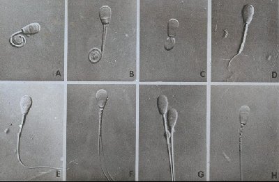
Photo of Sperm Defects: Photo: http://www.ansci.wisc.edu/jjp1/ansci_repro/lab/procedures/sperm/tertiary_abnormalities.jpg
Article:
http://www.cosmosmagazine.com/issues/2009/27/ (was obtained from magazine, need to be subscribed)
Sunday, May 31, 2009
What genes remember

Many geneticists now think that the behaviour of genes can be altered by experience and that these changes can be passed on to future generations.
In 1942 Conrad Waddington contrived the idea that an organism’s experience may cause genes to behave differently. He called this epigenetics. Until recently however it was assumed that the impact of epigenetics was confined to individual organisms and not passed on to their offspring. Scientists have become convinced of epigenetic inheritance, in which the behaviour of offspring is affected by the life experience of parents and furthermore these genes can extend to further generations although not lasting indefinitely. Epigenetic inheritance merely alters the ability of a gene to be expressed in offspring, but leaves the DNA, and the genes, intact. It can also be readily reversed and there is as yet little or no evidence that it persists for longer than a few generations.
Clear links between epigenetic inheritance and gene expression have not yet been found in humans. This would require multigenerational studies taking at least half a century. Fortunately there are historical records that provide striking indirect evidence of epigenetic inheritance surviving for at least two generations. The theory is still in early stages. However proof of it could help fill some of the gaps in evolutionary theory that creationists have exploited to bash Darwinism.
A Swap of Saliva Reveals Thyroid Cancer

The most common endocrine cancer is thyroid cancer, affecting approximately one percent of the population. Scientists from deCODE genetics, a bio-pharmaceutical company have discovered humans with the two single nucleotide polymorphism (SNPs) 9q22 and 14q13 located on their chromosome are at a higher risk of thyroid cancer. This was concluded after the analysis of 40,000 patients genomes from the companies database presenting that these two single letter variations in the human genome where in fact related to patients associated with an increased risk of the disease.
This information was earlier provided by a study carried out in 2009 by Gudmundsson et al. Information was gathered from European patients and found that the 9q22.33 allele is associated with low concentration of thyroxin (T-4) and high concentration of tri-iodothyronine (T-3) - releases more iodine within the hormones. However studies showed that both alleles are linked to producing low concentrations of thyroid stimulating hormone (TSH). Over expression of T3 and T4 lead to the increase in heart rate and blood flow to other organs, an increase in the carbohydrate and fat metabolism, increase in basal metabolic rate of almost all cells in the body as well as boosting protein synthesis. deCODE utilizes this information to benefit future patients that give the company a swap of their saliva for laboratory diagnostic testing.
Research using Aborigines makes maths breakthrough
Previously it was thought that human’s mathematic ability was due to the ability of language. A joint study between University College London and Melbourne University found that mathematical ability appears to be innate, or hard-wired into the human brain (Trouson, A 2009 ). It estimated from 4.3 to 10 percent of people have dyscalculia, a disability effecting ones mathematic ability. This disability is similar to dyslexia (Trouson, A 2009 ). The results published in the Washington journal “Proceeding of the National Academy of Science” and were based from 3 groups of aboriginal children. Two were non English speaking and the third was a group an English speaking from Melbourne. The non English speaking groups only had around four generic words for counting. It was found that both groups could do comparable maths.
The findings show that the treatment for dyscalculia may be misguided (Trouson, A 2009 ). The treated relies on language building and memory building. The findings show that it is a genetic or neurological disorder. In the report it shows that early detection maybe possible and that the teaching language for those who suffer should be used to reinforce basic mathematical principles. The findings are not a cure for the disability but can provide a new direction in which suffers can be treated
Trouson, A 2009 Research using Aborigines makes maths breakthrough, The Australian http://www.theaustralian.news.com.au/story/0,,24205070-12332,00.html
Written by Dominic Carroll, 42064084
A safer way to make stem-like cells

A research team in the U.S has found a new way to create stem-like cells using ordinary skin cells from patients. Their technique turns back the biological clock in human cells, making the cells behave like embryonic stem cells... minus the debate!
Four genes that can 'turn back time' in cells have been found by several different teams of researchers. The cells that have had their biological clock turned back are called induced pluripotent stem cells, or iPS cells for short. In theory, these iPS cells can be made from each individual patients' skin and used to produce transplants that have no risk of rejection by the patient.
Producing iPS cells is a difficult process. There have been a few different attempts at integrating the genetic material into the cells. One attempt used retroviruses (cells that inject their own genetic material into infected cells), another used the proteins produced by the four genes and acid to reprogram cells, but these methods all had drawbacks, Dr. Robert Lanza of Advanced Cell Technology Inc said.
Dr. Lanza's team uses a peptide to drag the human proteins into the cells to be transformed, a process that the AIDS virus also uses to get into infected cells.
According to Lanza, this method eliminates risks associated with genetic and chemical manipulation, and provides a potentially safe source of iPS cells for use in the clinic.
"Just add some proteins to some ordinary skin cells and you got patient-specific stem cells. It's the ultimate stem cell solution!"
Posted by: s4143590
Source: NewsDaily
~Understanding Cell Condensation~ by Lauren Crumlish
Scientists have discovered that cells, in a fashion similar to water vapour condensing into dew, are able to collect and focus subcellular organisms responsible for embryo development through utilisation of basic phase transition.
Discovered at the Marine Biological Laboratory by scientists in the 2008 Physiology course whilst studying C.elegans worms, scientists determed that subcellular 'P-granuales' are liquid droplets that shift between a dissolved and condensed states. when a single-celled embryo has been recently fertilised, the P-granuales are melting in the cell, much like water droplets do at high temperatures. Interestingly, just before the inital cell division, the same P-granules quickly solidify at the exterior of the cell where the temperature has decreased. The progenitor germ cell then develops where the P granules previously solidified. P granules are believed to be responsible for determining the 'germ cells' that eventually give rise to gametes.
http://www.sciencedaily.com/releases/2009/05/090521141204.htm
Nervous System May Be Culprit In Deadly Muscle Disease
Pompe disease is a rare inherited disorder that affects muscle function in infants. It is caused by a mutation in the GAA gene that prevents the enzyme acid alpha-glucosidase from breaking down glycogen. As a result, an accumulation of glycogen builds up in the body’s cells and impairs the muscles ability to function normally, particularly those of the heart and respiratory muscles. Children diagnosed with this disorder very rarely live beyond the age of two years old.


Left: Normal muscle cells
Right: affected muscle cells
In Pompe disease, glycogen accumulation causes the lysosomes to expand, damaging muscle cells. Glycogen leaks out of the cells, impairing muscle function.
Until a recent study, it was believed that Pompe disease was principally a muscle disease. In the Proceedings of the National Academy of Sciences investigation, researchers examined breathing in mice with Pompe disease and mice that were genetically engineered to produce GAA in muscle only and not in the central nervous system. It was found in both models that the phrenic nerve impulses (from the brain to the diaphragm via the spinal cord) to stimulate breathing were noticeably weaker than in normal mice. Furthermore, a detailed examination of a Pompe disease patient's nervous system showed a similar glycogen accumulation in the spinal cord as well as deficient neural output to the diaphragm.
These findings in the mouse models suggest that the disease may actually be caused by weakened signals from the brain to the diaphragm along the nervous system. Therefore treatments targeting the muscle alone may be inefficient. The study indicates that when treating children with Popme’s disease, not only do must the therapy be targeted to the muscle and heart, but also delivered to the nerve.
References:
University of Florida (2009, May 26). Nervous System May Be Culprit In Deadly Muscle Disease. ScienceDaily. Retrieved May 31, 2009, from http://www.sciencedaily.com /releases/2009/05/090525173444.htm
For more information on Pompe disease:
http://www.pompe.com/healthcare/overview/pc_eng_hc_overview_main.asp
http://ghr.nlm.nih.gov/condition=pompedisease
Saturday, May 30, 2009
Transgenic rodent sheds light on the evolution of language

The animal gene FOXP2 is involved in the development of language skills, however only humans contain key evolutionary changes in the gene. Scientists state that these changes are perhaps the reason why humans are the only animals able to communicate by multiple languages. Researches found a mutated copy of FOXP2 in a British family with a history of severe language disorders, they struggled to understand language and found it hard to speak.
A team of German researchers bred mice with the human version of FOXP2, attempting to discover the function of the gene. The team compared the traits of the transgenic mice with the traits of the normal mice and found differences in neuron production and ultrasonic calls. One German researcher states that the transgenic mice vocalisations are, “...at best similar to babies’ cries.” However they are still unsure as to how these results relate directly to human speech.
New Scientist http://www.newscientist.com/article/dn17206-human-speech-gene-gives-mouse-a-baritone-squeak.html
Junk DNA aren’t junk at all

Only 3% of human genomes are protein-coding DNA and scientist believes that the rest of the 97% are so-called ‘junk’ DNA which contains no genetic information. Recent research has found that tandem repeats, which are short stretches of DNA that are repeated, exactly influence the activity of the neighbouring genes.
Tandem repeats, a common example of ‘junk’ DNA, determine how tightly DNA is wrapped around a protein called nucleosomes which affects the level of activity of the gene. Also, scientist found out that these repeats are pretty unstable. They change their number of repeats frequently as DNAs are copied and these changes allow them to shift the gene’s activity effectively according to the changing environment, a key of survival to evolution.
Scientists test their theory by conducting a test using yeast cells, with one test-tube that has gene with tandem repeats and the other with the repeats removed. The results show that the test-tube with the repeats, scientists are able to identify the cells that have a great increased in gene activity and the test-tube without the repeat was unable to undergo fast evolution.
Hence, these ‘junk’ DNAs ability to adapt to changes very quickly gives organism an advantage in surviving countless evolution. Without them, cells would die as they would not be able to adapt to changes quickly enough. So, these ‘junk’ DNAs are exactly important life saviours in every organism.
Reference: VIB (the Flanders Institute for Biotechnology) (2009, May 30). Saved By Junk DNA: Vital Role In The Evolution Of Human Genome. ScienceDaily. Retrieved May 31, 2009, from http://www.sciencedaily.com /releases/2009/05/090528203730.htm
Posted by: RuBin Chua (41830143), Monday P2
New Therapy Substitutes Missing Protein in Those with Muscular Dystrophy
Duchenne muscular dystrophy is the most common and severe type of muscular dystrophy seen in children. This disease is a recessive X-linked trait, thus affecting mainly males, approximately 1 in 3500. The disease is characterized by the mutation of the gene DMD, which c odes for the protein dystrophin. This protein is an important part of the framework in the cell membrane that holds the muscle tissue together. Thus the symptoms of this disease are muscle weakness, and the rapid progression of muscle degeneration. Currently the only treatment for this disease is corticosteroids, which are minimally effective and have severe side effects.
 odes for the protein dystrophin. This protein is an important part of the framework in the cell membrane that holds the muscle tissue together. Thus the symptoms of this disease are muscle weakness, and the rapid progression of muscle degeneration. Currently the only treatment for this disease is corticosteroids, which are minimally effective and have severe side effects.
Cross-section of muscle (wild-type is normal muscle development)
(mdx is patient with muscular dystrophy)
However Professor James Ervasti from the University of Minnesota Medical School believe to have discovered a new therapy that shows potential to treat those with muscular dystrophy. The therapy includes injections of a protein called utrophin, which is a close relative of dystrophin, into the bloodstream. Researchers have currently used mouse models that were lacking in the protein dystrophin. These mice were injected with the protein utrophin that had been modified with a cell-penetrating tag known as TAT. The utrophin protein replaced the dystrophin in the cell membrane, and restored much of the strength of the muscle. This is the first study that has shown the viability of using this TAT-utrophin-based protein to treat the symptoms of muscular dystrophy. It is also thought that this therapy could assist in the treatment of cardiac muscle diseases that have resulted from the loss of dystrophin.
At the current time there are other therapies being developed using stem cell treatments, or delivering gene therapy to each individual muscle cell. Both these treatments show promise, but each has their limitations. From the mouse models it is shown that this method of protein replacement overcomes the limitations of both these methods. Firstly the TAT-utrophin protein spreads throughout the body efficiently and is able to penetrate the cell wall of all muscle cells so that it can replace the dystrophin in the cell membranes, and restore structure. Secondly as utrophin is produced naturally in small amount in every cell of the body, there is no risk of the immune system fighting off a foreign substance in the body.
This treatment of injecting utrophin proteins to replace the missing dystrophin in muscle cells has shown to be effective in improving the structure of muscle cells in mice, However before a it is developed as a drug for patients, the therapy will need to be trialled on much larger mammals and lastly humans. If this treatment does become a drug, it will not be a cure for muscular dystrophy, but a therapy that will need to be administered on a regular basis, to simulate the bodies natural process of producing dystrophin.
Posted by: Tanya McDonald 42048879
New Genetic Discovery may help people to regrow teeth

Researchers at the University of Rochester have discovered a method, which could lead to people being able to grow back several rows of teeth much like sharks. There is a gene researchers say that prevents people and most mammals from forming additional teeth. To combat this researchers bred mice lacking this gene, the mice gained backup teeth next to their molars much like sharks and other non-mammals have. Dr Songtao Shi of the University of Southern California School of Dentistry was quoted as saying “It’s exciting. We have a clue what to do,” in relation to the University of Rochersters discovery.
Issues such as gum disease resulting in tooth loss are a major problem and current treatment is insufficient. If scientist are able to discover what triggers new tooth growth they believe they may be able to switch that early-in-life process on again in adulthood so that people may be able to regrow missing teeth.
The breeding of these mice, although a great achievement in the quest towards the regrowth of teeth, has its setbacks. Mice bred without the gene called Osr2, had cleft pallets severe enough to kill. Scientists believe though that a better understanding of this gene will lead to being able to prevent this defect and come closer to the aim of regenerative teeth in humans.
Posted by 41789139
Link: http://www.nydailynews.com/news/us_world/2009/02/26/2009-02-26_new_genetic_discovery_may_help_people_to.htmlSaved By Junk DNA
 Researchers have found that DNA which was previously though to be ‘junk’, could play a vital role in the evolution of the genome.
It seems that tandem repeats influence the activity of neighbouring genes. How tightly local DNA wraps around nucleosomes is determined by these repeats. This packaging structure then dictates the extent to which genes can be activated. As these tandem repeats are very unstable the number present changes frequently during DNA replication. This changes the way DNA is packaged and activated.
These changes may allow organisms to tune the activity of their genes to match the environment. This is a vital principle for survival.
To test this idea scientists conducted a complex experiment testing tandem repeats in evolution using yeast cells as guinea pigs. It was found that when repeats were present near a gene it was “possible to select yeast mutants that show vastly increased activity of this gene”. When the tandem repeats were removed however this evolution was impossible.
As the researchers said “If this was the real world only cells with the repeats would be able to swiftly adapt to changes, thereby beating their repeat-less counterparts in the game of evolution. Their junk DNA saved their lives”.
Posted by 42053707
Link: http://www.medicalnewstoday.com/articles/151864.php
Evolution of Genetic Storage
In each human cell there is about 2m of genetic material packaged into our chromosomes. In order to fit such a large amount of genetic material into a cell, double-stranded human DNA is tightly wound using histones. Recently though, researchers at the University of Chicago discovered that single-celled aquatic algae called dinoflagellate have evolved an alternate way of packaging their DNA.
Packing of DNA is a difficult task due to the negative charge on the phosphate atom which must be neutralised to overcome electrostatic repulsion. Dinoflagellates have considerably more nuclear DNA that humans. In humans, positively-charged histone proteins bind DNA into nucleosomes. Dinoflagellates however, contain no histones or nucleosomes. So how do they condense their DNA into packages?
By using a high resolution scanning microprobe, researchers found that dinoflagellates had calcium and magnesium cations embedded in their DNA. This, in addition to the discovery that removal of calcium and magnesium cations caused an explosion of dinoflagellate chromosomes strongly suggested that these cations played a cricial role in the packing of DNA.
This leads to the question of how such evolution occurred in the first place. Did dinoflagellates initially have histones and over time, lost them? Or is this simply a case of convergent evolution, where alternate methods have evolved to perform the same function?
References:
- http://www.physorg.com/news138540908.html
posted by: 42022356
Friday, May 29, 2009
Genetic diseases a Thing of The Past
They say that everyone has a friend, relative or colleague who is personally affected by Breast Cancer. This fatal disease is the result of mutated or altered genes. Instead of regulating the growth of breast tissue, it causes cells to uncontrollably divide, making more than the normal amount of cells in a specific area, it is these cells that form tumours. Tumours fall into two categories malignant or benign.
Benign means not being life-threatening, in medical term this means that it is noncancerous and therefore not a threat to life or the long-term health of the patient.
Malignant on the other hand means being cancerous with cells that invades tissues around the infected area and if left undetected or untreated may invade vital organs of the body for example the liver and inevitably cause death.
Breast Cancer in particular claims over 2 600 Australians every year, averaging seven people per-day. Breast cancer is well publicized although little is known by the public about where when and how it starts.
Due to prominent people like, Belinda Emmett, Jane McGrath, Lance Armstrong and Kylie Minouge who have all suffered from some form of cancer and in some cases lost their battle, research and information is becoming readily available to the general public. This type of publicity has helped to make us aware of the need for regular checks and a greater awareness of this life threatening disease that appears to be able to strike anyone at anytime.
Both males and females have a chance of getting this disease. However research has shown that there are groups of people who have a higher risks factor due to:
1. Two or more close relatives, on the same side of the family or with a direct relative eg: mother or father, who have either had/ have prostate, breast or ovarian cancer.
2. Those who have the recently discovered mutated genes BRCA1 and BRCA2 (standing for BReast CAncer gene one and two) and whose effected relative is under 50.
People found with these genes have a 50-85% higher chance of getting Breast Cancer, than those who do not. The discovery of these mutations has assisted in the early detection in high risk patients and will ultimately have a significantly result in the prevention of this fatal disease. Research into and early intervention will significantly reduce the number of Breast Cancer cases, in people found with these genes.
Through new technologies in gene testing, patients have the opportunity to find out if they have these genes. A GP can take a blood sample and send it to a DNA analysis laboratory and within weeks patients should get the results. This relatively new technology was initially used to search for genetically inherited disorders, like Down Syndrome, Muscular Dystrophy and Hunting’s Disease, but as genetic scientists are now discovering more specific genes like BRCA1 and BRCA2 it is helping in the detection of patients who have high risk breast cancer factors.
BRCA1 and BRCA2 are located on chromosome 13 and 17 as displayed in the image to the right.
The possibility of gene testing and prevention of disease like breast cancer has been a stupendous break-though in medical science. Continued research developments is paramount in the continual battle to prevent genetic diseases, such as Breast Cancer, and other breakthroughs in medical science will continue as long as there is the passion and dedication of medical researchers, availability of funds and the will and determination to find ways to fight diseases and improve health and life throughout the world.
Benign means not being life-threatening, in medical term this means that it is noncancerous and therefore not a threat to life or the long-term health of the patient.
Malignant on the other hand means being cancerous with cells that invades tissues around the infected area and if left undetected or untreated may invade vital organs of the body for example the liver and inevitably cause death.
Breast Cancer in particular claims over 2 600 Australians every year, averaging seven people per-day. Breast cancer is well publicized although little is known by the public about where when and how it starts.
Due to prominent people like, Belinda Emmett, Jane McGrath, Lance Armstrong and Kylie Minouge who have all suffered from some form of cancer and in some cases lost their battle, research and information is becoming readily available to the general public. This type of publicity has helped to make us aware of the need for regular checks and a greater awareness of this life threatening disease that appears to be able to strike anyone at anytime.
Both males and females have a chance of getting this disease. However research has shown that there are groups of people who have a higher risks factor due to:
1. Two or more close relatives, on the same side of the family or with a direct relative eg: mother or father, who have either had/ have prostate, breast or ovarian cancer.
2. Those who have the recently discovered mutated genes BRCA1 and BRCA2 (standing for BReast CAncer gene one and two) and whose effected relative is under 50.
People found with these genes have a 50-85% higher chance of getting Breast Cancer, than those who do not. The discovery of these mutations has assisted in the early detection in high risk patients and will ultimately have a significantly result in the prevention of this fatal disease. Research into and early intervention will significantly reduce the number of Breast Cancer cases, in people found with these genes.
Through new technologies in gene testing, patients have the opportunity to find out if they have these genes. A GP can take a blood sample and send it to a DNA analysis laboratory and within weeks patients should get the results. This relatively new technology was initially used to
 search for genetically inherited disorders, like Down Syndrome, Muscular Dystrophy and Hunting’s Disease, but as genetic scientists are now discovering more specific genes like BRCA1 and BRCA2 it is helping in the detection of patients who have high risk breast cancer factors.
BRCA1 and BRCA2 are located on chromosome 13 and 17 as displayed in the image to the right.
The possibility of gene testing and prevention of disease like breast cancer has been a stupendous break-though in medical science. Continued research developments is paramount in the continual battle to prevent genetic diseases, such as Breast Cancer, and other breakthroughs in medical science will continue as long as there is the passion and dedication of medical researchers, availability of funds and the will and determination to find ways to fight diseases and improve health and life throughout the world.
Thursday, May 28, 2009
Zebrafish Provide Model For Cancerous Melanoma In Humans
nside the cell, signals are delivered that direct the cell on whether to divide, migrate or even die. Cancer often results when the molecules that relay these signals become mutated so that they do not function normally. In cancer, cells divide, grow and migrate when they should not, therefore resulting in an aggressive disease that can spread throughout the body. A key molecule in this signaling pathway is RAS, and mutations in it are known to lead to cancer. In some cases, the type of RAS mutation is a predictor of a patient's response to treatment and their overall prognosis.
Therefore, scientists at the University of Manchester in England and the University Hospital Zürich in Switzerland generated several zebrafish with changes in RAS or other RAS-regulated proteins to create a useful model that can be used to study and understand human melanoma. Zebrafish are a useful tool to understand human disease because they are small, transparent, and easy to propagate and maintain. Tumors created from the pigmented cells of zebrafish, known as melanocytes, are easy to see against their thin, light colored bodies.
The research team notes that these fish may be a useful experimental tool for human disease. Many of the changes they made caused melanoma in the zebrafish, indicating that zebrafish respond similarly to changes in these signals as do humans. Zebrafish that were born from the original mutant fish displayed abnormal growth of their melanocytes, reminiscent of familial atypical mole and melanoma syndrome (FAMM) seen in humans. By producing other signaling molecules in the mutant fish, the researchers were able to identify a pathway that reduced the effects of RAS mutations on melanoma progression in zebrafish.
The report titled "Dissecting the roles of Raf- and PI3K-signalling pathways in melanoma formation and progression in a zebrafish model" was written by Christina Michailidou, Mary Jones, Paul Walker, Amanda Kelly, and Adam Hurlstone at the University of Manchester and Jivko Kamarashev at University Hospital Zürich. The study is published in the July/August issue of the new research journal, Disease Models & Mechanisms (DMM), published by The Company of Biologists, a non-profit based in Cambridge, UK.
Subscribe to:
Comments (Atom)